November 2018
Thank you for your willingness to help with this project!
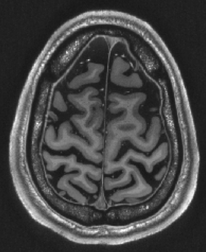
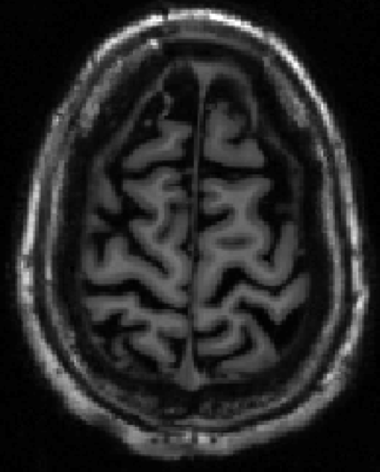
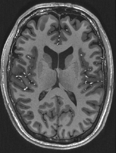
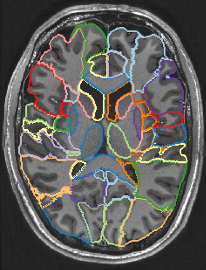
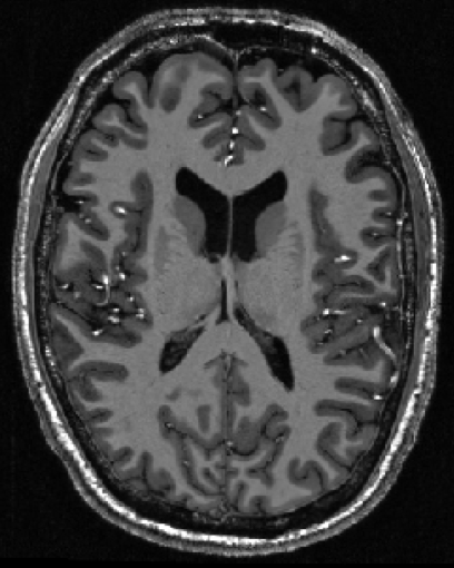
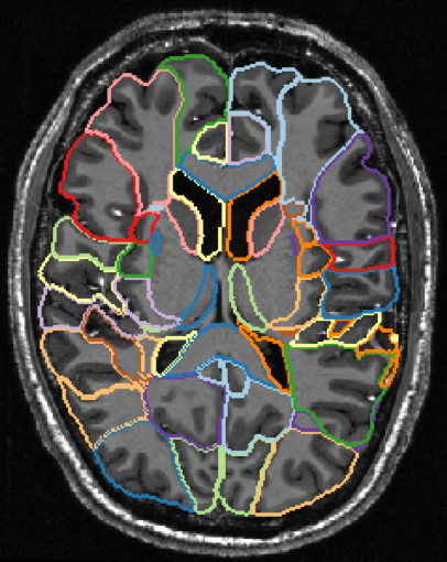
In the following, you will find sets of brain images to review. The images are from a single subject studied with varying parameters. The segmentation strategies also differ between the images.
With your help, I would like to find out if some combinations of acquisition parameters and processing methods are better than others, leading to higher segmentation quality as rated visually.
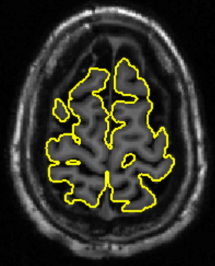
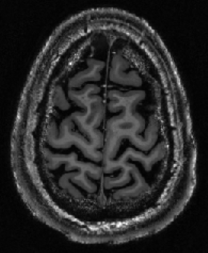
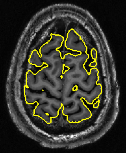
Each image is shown twice, once with and once without the segmentation boundaries superimposed. Please disregard the varying image quality and assess just the segmentation quality.
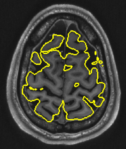
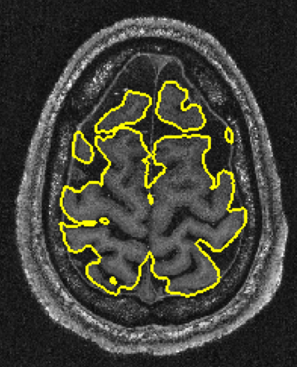
Four images are shown. The yellow line is supposed to delineate the parenchyma of the brain. Please order the images by segmentation quality from best to worst (e.g. D-A-C-B). A free-text comment on how you decided the ranking would be appreciated.

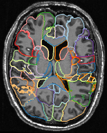
Three images are shown. They have been segmented into 95 regions. The coloured lines indicate the boundaries of the subset of regions encountered in this section. Please order the images by segmentation quality from best to worst. A free-text comment on how you decided the ranking would be appreciated.

Please note your thoughts about the experience of evaluating segmentations in this fashion. Would you be willing to participate again? What changes to the questionnaire design would you suggest?
Please send your answers by email to gusjohfrbm@student.gu.se.